










Cell Structure and Organisation
SEAB syllabus topic 1: the structures of plant and animal cells, membrane systems and organelles, how animal and plant cells compare, and how specialised cells are adapted, with the why, the practical, the traps and the exam checks.
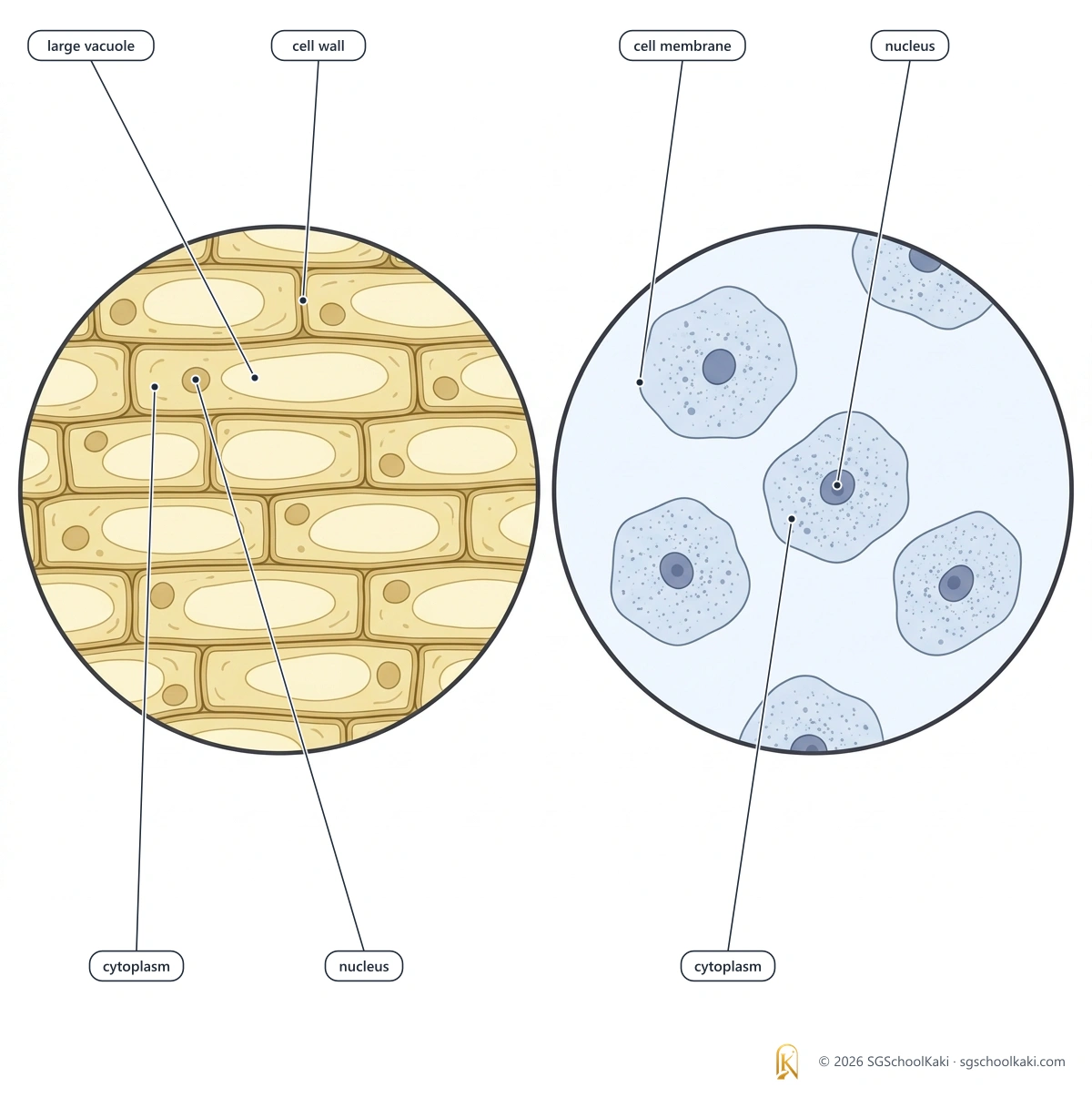

A typical plant cell has a cell wall and a large permanent central vacuole, and it has chloroplasts in the green parts of the plant; an animal cell has none of these.
Mitochondria are the site of aerobic respiration and ribosomes are the site of protein synthesis.
A specialised cell has a structure that is adapted to its function.
Cell structures seen under the light microscope
identify and state the functions of the following cell structures (including organelles) of typical plant and animal cells from diagrams, light micrographs and as seen under the light microscope using prepared slides and fresh material treated with an appropriate temporary staining technique: • cell wall • cell membrane • cytoplasm • nucleus • cell vacuoles (large, sap-filled in plant cells, small, temporary in animal cells) • chloroplasts
Each part of a cell has its own job, and together the parts keep the cell alive.
- Cell
- Cell: the basic unit of structure and function of living things
- Organelle
- Organelle: a structure inside a cell that has a particular job, for example the nucleus or a mitochondrion
- Cell membrane
- Cell membrane: the thin layer around the cytoplasm, which is partially permeable and so controls what moves into and out of the cell
- Cell wall
- Cell wall: a layer of cellulose outside the cell membrane of a plant cell, which is fully permeable and gives the cell its shape
- Cytoplasm
- Cytoplasm: the jelly-like substance that fills the cell, in which the organelles are found
- Nucleus
- Nucleus: the organelle that contains the DNA of the cell and controls the activities of the cell
- Vacuole
- Vacuole: a fluid-filled space in the cytoplasm; a plant cell has one large central vacuole and an animal cell has several small, temporary vacuoles
- Cell sap
- Cell sap: the solution in the large central vacuole of a plant cell, which contains water, sugars, amino acids and mineral salts
- Chloroplast
- Chloroplast: a green organelle that contains chlorophyll and is the site of photosynthesis
- Light microscope
- Light microscope: a microscope that uses light and glass lenses to magnify a specimen, and it can be used to look at living cells
Cell structures and their functions
| Structure | Description | Function | Seen with a light microscope? |
|---|---|---|---|
| Cell wall (plant cells) | The cell wall is a layer of cellulose outside the cell membrane of a plant cell. | The cell wall is fully permeable, so water and dissolved substances pass through it, and it gives the cell its shape and support. | The cell wall can be seen under a light microscope. |
| Cell membrane | The cell membrane is a very thin layer around the cytoplasm, just inside the cell wall in a plant cell. | The cell membrane is partially permeable, and it controls the movement of substances into and out of the cell. | The cell membrane is seen under a light microscope only as the boundary of the cell, because it is too thin to show any detail. |
| Cytoplasm | The cytoplasm is a jelly-like substance, made mostly of water, that fills the cell and contains the organelles. | Many of the chemical reactions of the cell take place in the cytoplasm, and anaerobic respiration takes place in the cytoplasm. | The cytoplasm can be seen under a light microscope. |
| Nucleus | The nucleus is a large, round organelle that contains the DNA of the cell. | The nucleus controls the activities of the cell. | The nucleus can be seen under a light microscope, and a stain makes it easier to see. |
| Nuclear envelope | The nuclear envelope is the membrane that surrounds the nucleus. | The nuclear envelope separates the nucleus from the cytoplasm. | The outline of the nucleus can be seen under a light microscope, but the structure of the nuclear envelope is seen only in electron micrographs. |
| Vacuole (plant cell) | A plant cell has one large central vacuole, which is filled with cell sap. | The large central vacuole stores cell sap, which is a solution of water, sugars, amino acids and mineral salts. | The large central vacuole of a plant cell can be seen under a light microscope. |
| Vacuole (animal cell) | An animal cell has several small vacuoles, and each one is temporary. | The small vacuoles of an animal cell store food and water. | The small vacuoles of an animal cell are small and temporary, so they are hard to see clearly under a light microscope. |
| Chloroplast (plant cells) | Chloroplasts are green organelles that contain the green pigment chlorophyll, and they are found in many cells of the green parts of a plant, such as the palisade and spongy mesophyll cells, but not in root cells or leaf epidermal cells. | Chloroplasts are the site of photosynthesis, in which light energy is used to make glucose. | Chloroplasts can be seen under a light microscope as small green discs. |
The cell wall is fully permeable and the cell membrane is partially permeable, so if a question asks what controls the movement of substances into and out of a cell, the answer is the cell membrane.
Some textbooks call the cell membrane selectively permeable, but for SEAB answers, write partially permeable.
Seeing cells: light microscope and electron microscope
| Feature | Light microscope | Electron microscope |
|---|---|---|
| What it uses | A light microscope uses light and glass lenses. | An electron microscope uses a beam of electrons. |
| Resolution | A light microscope has a lower resolution, so it cannot show very small organelles such as ribosomes. | An electron microscope has a much higher resolution, so it shows the small organelles and the fine structure of the cell. |
| The specimen | Living cells and fresh material can be viewed with a light microscope. | The specimen must be dead to be viewed with an electron microscope. |
| Where you meet it | The cell wall, cell membrane, cytoplasm, nucleus, vacuoles and chloroplasts are identified from light micrographs and from slides seen under the light microscope. | The endoplasmic reticulum, Golgi body, mitochondria and ribosomes are identified from electron micrographs. |
🧪 Practical skills: looking at cells under a light microscope
Method
- 1Peel a thin layer of epidermis from the inside of an onion scale leaf with forceps, and lay it flat in a drop of iodine solution on a clean slide.
- 2For animal cells, gently scrape the inside of your cheek with a clean cotton bud, smear the cells on a drop of methylene blue on a clean slide, and put the cotton bud in disinfectant.
- 3Lower a coverslip onto the specimen at an angle, using a mounted needle, so that no air bubbles are trapped.
- 4Start with the low-power objective lens, focus with the coarse focus and then the fine focus, and only then change to a higher-power objective lens and focus again with the fine focus only.
- 5For chloroplasts, mount a fresh leaf of Hydrilla in a drop of water, with no stain, and look at it in the same way.
What you see
Onion epidermal cell
An onion epidermal cell shows a cell wall, cytoplasm, a nucleus and a large vacuole (the cell membrane lies just inside the cell wall but cannot be seen separately), and it has no chloroplasts because the onion bulb grows underground and receives no light.
Cheek cell
A cheek cell is rounded or irregular in shape, and it shows a cell membrane, cytoplasm and a nucleus, but no cell wall and no chloroplasts.
Hydrilla leaf cell
A Hydrilla leaf cell shows a cell wall and many green chloroplasts in the cytoplasm.
Working out the magnification
- 1Magnification = size of the image ÷ size of the real object, and both sizes must be in the same unit.
- 2For example, if a drawing of a cell is 60 mm long and the cell is really 0.3 mm long, the magnification is 60 ÷ 0.3 = ×200.
- 3Magnification has no unit, so write it as ×200.
In a biological drawing, use a sharp pencil, draw single clear lines, do not shade or colour, and draw the label lines with a ruler so that each one touches the structure it names. supports K325-1(a)
Depth: why and how
- Why is a stain used on onion cells and cheek cells? Most cells are colourless and transparent, so a stain makes structures such as the nucleus easier to see.
- Why is the coverslip lowered at an angle? This pushes the air out, and air bubbles look like dark rings that can be mistaken for cells.
- Why do onion bulb cells have no chloroplasts? The bulb grows underground, so no light reaches it and its cells do not carry out photosynthesis; many cells in the green parts of a plant have chloroplasts.
- Why is the vacuole of a plant cell easier to see than the vacuoles of an animal cell? The plant cell has one large central vacuole, whereas the vacuoles of an animal cell are small and temporary.
- Cross-link to Movement of substances: the cell membrane is partially permeable, which is why osmosis can take place across it.
- Cross-link to Respiration in humans: aerobic respiration takes place in the mitochondria, and anaerobic respiration takes place in the cytoplasm.
- When the large central vacuole is full of cell sap, it pushes the cytoplasm against the cell wall and the plant cell is firm, which is called turgid. supports K325-2(b)
Common mistakes
The cell wall controls what enters and leaves the cell.
The cell wall is fully permeable, and the partially permeable cell membrane controls what enters and leaves the cell.
Animal cells have no vacuoles.
Animal cells have several small, temporary vacuoles, and plant cells have one large, permanent central vacuole.
All plant cells contain chloroplasts.
Many plant cells in the green parts of a plant contain chloroplasts, but root cells do not, because no light reaches them.
The cell wall and the cell membrane are the same thing.
The cell wall is made of cellulose and is outside the cell membrane, and plant cells have one but animal cells do not.
Wise Monkeys Can Never Value Cheese: Wall of cellulose gives plant cells their shape, Membrane controls what enters and leaves the cell, Cytoplasm is where many reactions happen, Nucleus controls the cell, Vacuole stores cell sap or food and water, Chloroplast is the site of photosynthesis.
Decode key: W is the cell wall, which plant cells have, M is the cell membrane, the first C is the cytoplasm, N is the nucleus, V is the vacuole, and the last C is the chloroplast, which plant cells have.The cell membrane is partially permeable and controls what enters and leaves the cell, whereas the cell wall of a plant cell is fully permeable and gives the cell its shape.
Exam check
Q1 Which part of a plant cell is made of cellulose and is fully permeable?
- A Cell membrane
- B Cell wall
- C Cytoplasm
- D Nucleus
Show answer
MCQ answer key B (Cell wall): the cell wall is made of cellulose, it is fully permeable, and it gives the plant cell its shape.
Q2 Which structure controls the activities of a cell?
- A Cytoplasm
- B Vacuole
- C Chloroplast
- D Nucleus
Show answer
MCQ answer key D (Nucleus): the nucleus contains the DNA of the cell and controls the activities of the cell.
Q3 Which structure is not found in a human cheek cell?
- A Cell wall
- B Cell membrane
- C Nucleus
- D Cytoplasm
Show answer
MCQ answer key A (Cell wall): an animal cell such as a cheek cell has a cell membrane, cytoplasm and a nucleus, but it has no cell wall.
Structured A student mounts a fresh leaf of Hydrilla in water and looks at it under a light microscope.
- Name the organelle that gives the leaf cells their green colour. [1]
- State the function of this organelle. [1]
- State two differences between a Hydrilla leaf cell and a human cheek cell. [2]
- Explain why she lowers the coverslip at an angle. [1]
Total: 5 marks
Show mark scheme
(a) 1 mark
- Mark scheme: chloroplast (chloroplasts)
(b) 1 mark
- Mark scheme: it is the site of photosynthesis
(c) 2 marks
- Mark scheme: any two of: the leaf cell has a cell wall and the cheek cell does not
- Mark scheme: the leaf cell has chloroplasts and the cheek cell does not
- Mark scheme: the leaf cell has a large central vacuole and the cheek cell has only small, temporary vacuoles
(d) 1 mark
- Mark scheme: so that air bubbles are not trapped, because air bubbles look like cells and would make the specimen hard to see
You've read the free 25%. Unlock the rest of Topic 1.
Still to read in this topic:
- Membrane systems and organelles
- Comparing animal and plant cells
- Specialised cells
$9 one-time · all 14 topics · no subscription
Less than a fast-food meal.
Yours for the whole O-Level.
Premium members get all 14 topics included.
Notes version 1.0 · Last updated 30 Sept 2026 · Topic 1 of 14