










Respiration in Humans
SEAB syllabus topic 6: the gas exchange system, breathing, tobacco smoke, aerobic and anaerobic respiration and oxygen debt, with the why, the practical, the traps and the exam checks.
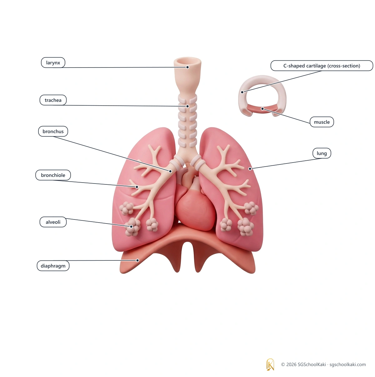

Breathing moves air into and out of the lungs by changing the volume of the thorax.
Gaseous exchange takes place by diffusion across the wall of the alveolus and the wall of the capillary.
Respiration releases energy from glucose in the cells, with oxygen in aerobic respiration and without oxygen in anaerobic respiration.
Structures of the gas exchange system
identify the larynx, trachea, bronchi, bronchioles, alveoli and associated capillaries and state their functions in human gaseous exchange
Air that is breathed in travels through the nasal passage, pharynx, larynx, trachea, bronchi and bronchioles to the alveoli, where gaseous exchange takes place between the air and the blood in the capillaries.
- Gaseous exchange
- Gaseous exchange: the movement of oxygen from the air into the blood and of carbon dioxide from the blood into the air, by diffusion, in the alveoli
- Larynx
- Larynx: the voice box at the top of the trachea; air passing over the vocal cords in the larynx produces sound
- Trachea
- Trachea: the windpipe, a tube that carries air from the larynx to the bronchi; C-shaped rings of cartilage in its wall keep it open
- Bronchus
- Bronchus: one of the two tubes into which the trachea divides, one leading to each lung; the plural is bronchi
- Bronchiole
- Bronchiole: a narrow tube formed by the repeated branching of a bronchus; each bronchiole ends in a cluster of alveoli
- Alveolus
- Alveolus: a tiny air sac at the end of a bronchiole, where oxygen and carbon dioxide are exchanged between the air and the blood; the plural is alveoli
- Capillary
- Capillary: a very narrow blood vessel with a wall one cell thick; the capillaries around the alveoli bring blood to the alveoli and carry blood away from them
The route air takes
- 1Air enters through the nasal passage, where hairs and mucus trap dust and the lining warms and moistens the air.
- 2Air passes through the pharynx, which is the throat, to the larynx.
- 3Air passes through the larynx and into the trachea.
- 4The trachea divides into two bronchi, one leading to each lung.
- 5Each bronchus divides many times to form bronchioles.
- 6Each bronchiole ends in a cluster of alveoli, where gaseous exchange takes place.
Structures and their functions
| Structure | Feature | Function in gaseous exchange |
|---|---|---|
| Nasal passage | The nasal passage is lined with hairs and mucus. | The hairs and mucus trap dust, and the lining warms and moistens the air that passes through. |
| Larynx | The larynx is the voice box, at the top of the trachea. | The larynx is part of the air passage, so air passes through it on its way to the trachea. |
| Trachea | The wall of the trachea contains C-shaped rings of cartilage, and its inner lining has ciliated cells and mucus-secreting cells. | The trachea carries air to the bronchi, and the cartilage rings keep it open so that air can flow freely. |
| Bronchi | The two bronchi are the branches of the trachea, and each one leads to a lung. | The bronchi carry air into the lungs. |
| Bronchioles | The bronchioles are narrow tubes formed by the repeated branching of the bronchi. | The bronchioles carry air to the alveoli. |
| Alveoli | The alveoli are tiny air sacs, grouped in clusters at the ends of the bronchioles. | Oxygen from the air diffuses across the wall of an alveolus into the blood, and carbon dioxide from the blood diffuses across the wall into the air in the alveolus. |
| Capillaries | The capillaries form a network around each alveolus, and their walls are one cell thick. | The capillaries bring blood that is low in oxygen and high in carbon dioxide to the alveoli, and they carry the blood, which is now rich in oxygen, away from the alveoli. |
Keeping the airways clean and open
Ciliated cells
Ciliated cells line the trachea, bronchi and larger bronchioles, and their cilia sweep mucus, with the dust and pathogens trapped in it, up towards the throat, where it is swallowed. supports K325-6(b)
Mucus-secreting cells
Mucus-secreting cells in the lining of the airways make mucus, which traps dust and other foreign particles from the air. supports K325-6(b)
C-shaped rings of cartilage
The C-shaped rings of cartilage support the trachea and stop it from collapsing, so the airway stays open. supports K325-6(a)
Stomach acid
The acid in the stomach kills most of the pathogens that are swallowed with the mucus. supports K325-6(b)
Depth: why and how
- Why does the trachea have C-shaped rings of cartilage? The rings are stiff, so they keep the trachea open when the air pressure inside it falls during inspiration, and the gap at the back of each ring lets the oesophagus expand when food is swallowed.
- Why do the airways branch into so many bronchioles? The repeated branching carries air close to a very large number of alveoli, which gives a large surface area for gaseous exchange.
- Why is a network of capillaries wrapped around each alveolus? The blood brings carbon dioxide and takes oxygen away, so the concentration gradients of both gases are kept steep.
- What happens to dust that gets past the hairs in the nose? The mucus in the trachea and bronchi traps it, and the cilia sweep the mucus up to the throat to be swallowed.
- Cross-link to Transport in humans: the pulmonary artery carries blood that is low in oxygen from the heart to the lungs, and the pulmonary vein carries blood that is rich in oxygen back to the heart.
- Cross-link to Movement of substances: oxygen and carbon dioxide cross the wall of the alveolus by diffusion, which is a passive process.
Common mistakes
The trachea carries food to the stomach.
The trachea carries air to the bronchi, and food travels down the oesophagus.
Gaseous exchange takes place in the bronchi.
Gaseous exchange takes place in the alveoli, and the trachea, bronchi and bronchioles only carry air to them.
The lungs are hollow bags full of air.
The lungs contain millions of alveoli, grouped in clusters, which gives them a large surface area.
Blood flows through the alveoli.
Blood flows through the capillaries around the alveoli, and gases diffuse between the air in the alveoli and the blood.
Lions Travel Barefoot, Bravely Alone: Larynx, Trachea, Bronchi, Bronchioles, Alveoli, in the order that air travels.
The first B is for the bronchi, which are the wider tubes, and the second B is for the bronchioles, which are the narrower tubes.Air is carried to the alveoli through the larynx, trachea, bronchi and bronchioles, and gaseous exchange takes place only at the alveoli.
Exam check
Q1 Which structure is the site of gaseous exchange?
- A Trachea
- B Bronchus
- C Alveolus
- D Bronchiole
Show answer
MCQ answer key C (Alveolus): gaseous exchange takes place across the thin walls of the alveoli, whereas the trachea, bronchi and bronchioles only carry air.
Q2 Which sequence shows the order in which air passes through the structures?
- A Larynx, bronchi, trachea, bronchioles, alveoli
- B Larynx, trachea, bronchi, bronchioles, alveoli
- C Trachea, larynx, bronchi, bronchioles, alveoli
- D Larynx, trachea, bronchioles, bronchi, alveoli
Show answer
MCQ answer key B (Larynx, trachea, bronchi, bronchioles, alveoli): the trachea divides into bronchi, each bronchus divides into bronchioles, and the bronchioles end in alveoli.
Q3 What is the job of the capillaries around an alveolus?
- A To make mucus that traps dust
- B To sweep dust away from the alveolus
- C To let air pass into the alveolus
- D To bring blood low in oxygen to the alveolus and carry oxygen away in the blood
Show answer
MCQ answer key D (To bring blood low in oxygen to the alveolus and carry oxygen away in the blood): the capillaries bring blood with less oxygen to the alveolus and carry the oxygen-rich blood away, which keeps the concentration gradients steep.
Structured Air moves through the airways to the lungs.
- Name the tube that carries air from the larynx to the bronchi. [1]
- State how the trachea is kept open. [1]
- State one function of the capillaries around the alveoli. [1]
Total: 3 marks
Show mark scheme
(a) 1 mark
- Mark scheme: trachea
(b) 1 mark
- Mark scheme: the wall contains C-shaped rings of cartilage
(c) 1 mark
- Mark scheme: any one of: bring blood that is low in oxygen to the alveoli, OR carry oxygen away from the alveoli in the blood, OR bring carbon dioxide to the alveoli
Breathing
describe the process of breathing and the role of cilia, diaphragm, ribs and internal and external intercostal muscles
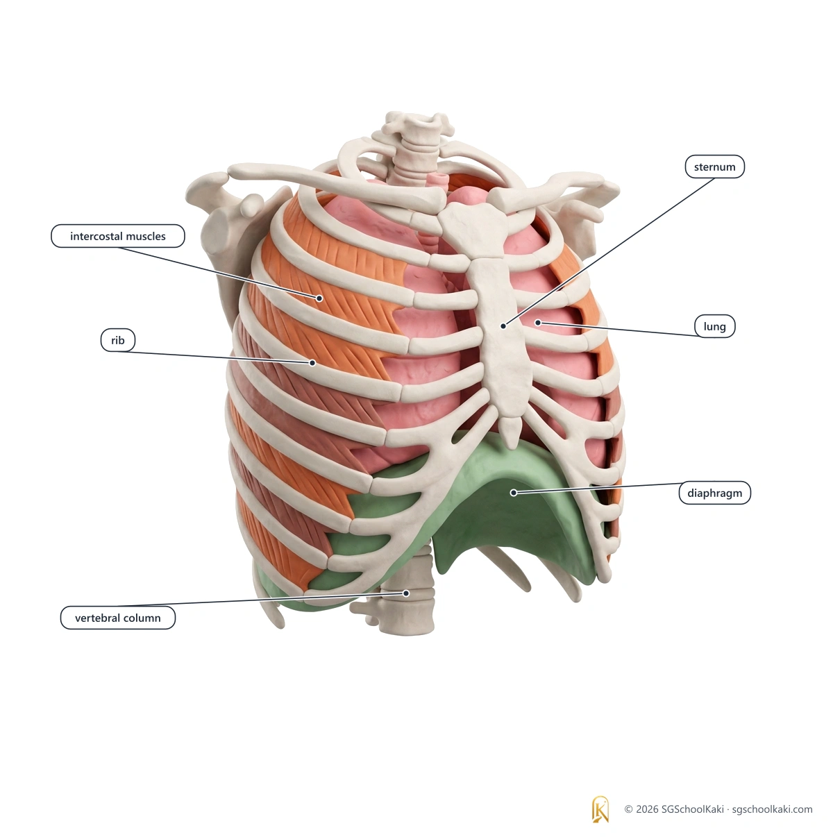
Breathing is the movement of air into and out of the lungs, caused by changes in the volume of the thorax, which change the air pressure in the lungs.
- Breathing
- Breathing: the movement of air into and out of the lungs; another word for it is ventilation
- Inspiration
- Inspiration: breathing in, when air moves into the lungs; another word for it is inhalation
- Expiration
- Expiration: breathing out, when air moves out of the lungs; another word for it is exhalation
- Thorax
- Thorax: the chest, which is enclosed by the rib cage and separated from the abdomen by the diaphragm supports K325-6(b)
- Diaphragm
- Diaphragm: a sheet of muscle below the lungs that separates the thorax from the abdomen
- Intercostal muscles
- Intercostal muscles: muscles between the ribs; the external intercostal muscles are on the outside and the internal intercostal muscles are on the inside
- Antagonistic muscles
- Antagonistic muscles: two muscles that work against each other, so that when one contracts the other relaxes supports K325-6(b)
- Cilia
- Cilia: tiny hair-like structures on the surface of ciliated cells, which beat in a sweeping movement
The rib cage
The rib cage
The rib cage is made of the ribs, the sternum at the front and the spine at the back, and it protects the lungs and the heart. supports K325-6(b)
How the ribs are joined
The ribs are attached to the spine at the back and, for most of the ribs, to the sternum at the front. supports K325-6(b)
What changes when we breathe
| What changes | Inspiration (breathing in) | Expiration (breathing out) |
|---|---|---|
| Diaphragm | The diaphragm contracts and flattens. | The diaphragm relaxes and curves upwards into a dome shape. |
| External intercostal muscles | The external intercostal muscles contract. | The external intercostal muscles relax. |
| Internal intercostal muscles | The internal intercostal muscles relax. | The internal intercostal muscles contract. |
| Rib cage | The rib cage moves upwards and outwards. | The rib cage moves downwards and inwards. |
| Volume of the thorax | The volume of the thorax increases. | The volume of the thorax decreases. |
| Air pressure in the lungs | The air pressure in the lungs becomes lower than the air pressure outside, so air moves into the lungs. | The air pressure in the lungs becomes higher than the air pressure outside, so air moves out of the lungs. |
The external and the internal intercostal muscles are an antagonistic pair, so when one set contracts the other set relaxes. supports K325-6(b)
Step by step: breathing in and breathing out
Breathing in
- 1The diaphragm contracts and flattens, the external intercostal muscles contract and the internal intercostal muscles relax.
- 2The rib cage moves upwards and outwards.
- 3The volume of the thorax increases.
- 4The air pressure in the lungs falls below the air pressure outside.
- 5Air moves into the lungs from outside.
Breathing out
- 1The diaphragm relaxes and curves upwards, the external intercostal muscles relax and the internal intercostal muscles contract.
- 2The rib cage moves downwards and inwards.
- 3The volume of the thorax decreases.
- 4The air pressure in the lungs rises above the air pressure outside.
- 5Air moves out of the lungs to the outside.
Cilia and mucus: keeping the air clean
Role of cilia
The cilia in the lining of the trachea, bronchi and larger bronchioles beat to sweep mucus, with the dust and pathogens trapped in it, up towards the throat, so the air that reaches the alveoli is cleaner.
Role of mucus
Mucus made by the cells in the lining of the airways traps dust and pathogens, so that they can be swept away by the cilia. supports K325-6(b)
Depth: why and how
- Why does air move into the lungs when the thorax gets bigger? A larger volume gives a lower pressure in the lungs, so the air pressure outside is higher, and air moves from the higher pressure to the lower pressure.
- Why must the diaphragm and the intercostal muscles keep working? The lungs have no muscles of their own to expand them, so they can only be moved by the changes in the volume of the thorax that these muscles cause.
- How does breathing help gaseous exchange? Breathing brings fresh air into the alveoli and removes air that contains more carbon dioxide, so the concentration gradients for both gases stay steep.
- Cross-link to Movement of substances: breathing does not move the gases across the wall of the alveolus, because that is done by diffusion; breathing keeps the concentration gradients steep so that diffusion can continue.
- Cross-link to Respiration: breathing takes in the oxygen that cells need for aerobic respiration and removes the carbon dioxide that respiration produces.
- In quiet breathing, expiration is mostly passive, because the elastic tissue in the lungs recoils and pushes air out, and the internal intercostal muscles contract mainly when a person breathes out forcefully. For SEAB answers, write that the internal intercostal muscles contract and the ribs move down and in. supports K325-6(b)
Common mistakes
The lungs pull themselves bigger to suck air in.
The lungs have no muscles of their own, and the volume of the thorax changes because the diaphragm and the intercostal muscles move.
During inspiration the diaphragm relaxes and moves up.
During inspiration the diaphragm contracts and flattens.
Breathing is the same as respiration.
Breathing is the movement of air into and out of the lungs, but respiration is the release of energy from glucose in cells.
The ribs move down when we breathe in.
During inspiration the rib cage moves upwards and outwards.
Air enters the lungs because the pressure outside becomes lower.
Air enters the lungs because the air pressure in the lungs becomes lower than the air pressure outside.
Breathe IN and the changes DRIP in: Diaphragm contracts and flattens, Ribs move up and out because the external intercostal muscles contract, Inside the thorax the volume increases, Pressure in the lungs decreases so air rushes in; breathing out reverses all four, and the internal intercostal muscles contract.
The letters D, R, I and P stand for the four changes; the diaphragm and the external intercostal muscles are the two muscles that contract, and the pressure that decreases is the air pressure in the lungs.When you breathe in, the thorax gets bigger and the air pressure in the lungs falls, so air moves in; when you breathe out, the thorax gets smaller and the air pressure in the lungs rises, so air moves out.
Exam check
Q1 What happens to the diaphragm and the rib cage during inspiration?
- A The diaphragm relaxes and the rib cage moves downwards and inwards
- B The diaphragm contracts and the rib cage moves downwards and inwards
- C The diaphragm contracts and flattens, and the rib cage moves upwards and outwards
- D The diaphragm relaxes and the rib cage moves upwards and outwards
Show answer
MCQ answer key C (The diaphragm contracts and flattens, and the rib cage moves upwards and outwards): during inspiration the diaphragm contracts and flattens, and the rib cage moves upwards and outwards, which increases the volume of the thorax.
Q2 Which change causes air to move out of the lungs during expiration?
- A The volume of the thorax increases, so the air pressure in the lungs falls
- B The volume of the thorax decreases, so the air pressure in the lungs rises
- C The volume of the thorax decreases, so the air pressure in the lungs falls
- D The volume of the thorax increases, so the air pressure in the lungs rises
Show answer
MCQ answer key B (The volume of the thorax decreases, so the air pressure in the lungs rises): a smaller thorax raises the air pressure in the lungs above the air pressure outside, so air moves out.
Q3 Which statement about the external and the internal intercostal muscles is correct?
- A They are antagonistic, so when the external set contracts the internal set relaxes
- B They both contract at the same time during inspiration
- C They are found inside the lungs
- D They both relax during inspiration
Show answer
MCQ answer key A (They are antagonistic, so when the external set contracts the internal set relaxes): the external and the internal intercostal muscles are antagonistic, so when the external intercostal muscles contract during inspiration the internal intercostal muscles relax.
Structured A student breathes in deeply.
- State what happens to the diaphragm when she breathes in. [1]
- Explain why air moves into her lungs. [3]
- Describe the role of the cilia in her trachea. [2]
Total: 6 marks
Show mark scheme
(a) 1 mark
- Mark scheme: the diaphragm contracts and flattens
(b) 3 marks
- Mark scheme: the volume of the thorax increases
- Mark scheme: so the air pressure in the lungs becomes lower than the air pressure outside
- Mark scheme: air moves from the higher air pressure outside into the lungs
(c) 2 marks
- Mark scheme: the cilia sweep mucus, containing trapped dust and pathogens, upwards towards the throat
- Mark scheme: so that the mucus can be swallowed and the air reaching the alveoli is cleaner
You've read the free 40%. Unlock the rest of Topic 6.
Still to read in this topic:
- How the alveolus is suited to its function
- Tobacco smoke and health
- Aerobic respiration
- Anaerobic respiration
- Vigorous exercise and oxygen debt
$9 one-time · all 14 topics · no subscription
Less than a fast-food meal.
Yours for the whole O-Level.
Premium members get all 14 topics included.
Notes version 1.0 · Last updated 30 Sept 2026 · Topic 6 of 14