










Homeostasis, Co-ordination and Response in Humans
SEAB syllabus topic 8: how the body keeps its internal environment constant, and how the nervous system and hormones co-ordinate the response, with the why, the traps and the exam checks.

This topic covers homeostasis and negative feedback, hormones, the control of the blood glucose concentration and diabetes, osmoregulation and ADH, the skin and the control of body temperature, the nervous system and reflex actions, and the human eye.
The nervous system part of the topic covers the CNS and the PNS, neurones, reflex actions, the structure and the focusing of the human eye, and a comparison of the nervous system with the endocrine system.
Negative feedback keeps the internal environment constant: a receptor detects a change and a corrective mechanism reverses it.
Hormones such as insulin and glucagon are carried by the blood to target organs, where they control the blood glucose concentration.
The nervous system uses receptors, neurones and effectors to produce a fast, co-ordinated response.
What is homeostasis?
define homeostasis as the maintenance of a constant internal environment
Homeostasis is the maintenance of a constant internal environment.
- Internal environment
- Internal environment: the blood and the tissue fluid that surround the cells of the body supports K325-8(a)
- Set point
- Set point: the normal value of a condition in the internal environment, such as a body temperature of about 37 °C supports K325-8(b)
- Constant
- Constant: kept within narrow limits around the set point, so the value rises and falls by a small amount and homeostasis brings it back supports K325-8(a)
- Homeostatic organs
- Homeostatic organs: the organs that take part in homeostasis include the skin, the brain, the pancreas, the liver and the kidneys supports K325-8(a)
What is kept constant, and why
| Condition that is controlled | Why it must be kept constant |
|---|---|
| Body temperature | Enzymes work best at their optimum temperature, so a large rise in body temperature can denature enzymes and a fall in body temperature slows down the reactions in the cells. |
| Water potential of the blood | If the water potential of the blood is too high or too low, water moves into or out of the cells by osmosis, and the cells can swell or shrink. |
| Blood pressure | The blood pressure must be high enough to push blood through the capillaries to the cells, but not so high that it damages the blood vessels. |
| Blood pH and carbon dioxide concentration | Enzymes work best at their optimum pH, and a high concentration of carbon dioxide in the blood makes the blood more acidic. |
| Blood glucose concentration | Cells need a supply of glucose for aerobic respiration, and a very high blood glucose concentration changes the water potential of the blood. |
| Water content of the urine | The kidneys change the water content of the urine to keep the water potential of the blood constant. |
In a definition of homeostasis, write 'a constant internal environment'; do not write that the body 'stays the same' or that the temperature 'does not change'.
Depth: why and how
- Why must the internal environment be kept constant? The cells work best in a narrow range of conditions, so a change in temperature, pH, water potential or glucose concentration would slow down or damage the cells.
- Why are the blood and the tissue fluid called the internal environment? They are in direct contact with the cells, so their condition decides the condition of the cells.
- Cross-link to Movement of substances: the water potential of the blood must be kept constant so that the cells do not gain or lose too much water by osmosis.
- Cross-link to Excretion in humans: the kidneys help to keep the water potential of the blood constant by changing the water content of the urine.
- Cross-link to Nutrition in humans: the liver stores glucose as glycogen, which helps to keep the blood glucose concentration constant.
Common mistakes
Homeostasis means that the conditions in the body do not change at all.
Homeostasis is the maintenance of a constant internal environment, so the conditions stay within narrow limits around the set point.
The internal environment is the inside of the stomach and the intestines.
The internal environment is the blood and the tissue fluid that surround the cells of the body.
Homeostasis controls body temperature and nothing else.
Homeostasis also controls the blood glucose concentration, the water potential of the blood, the blood pressure and the blood pH.
Homeostasis keeps the internal environment constant, and that keeps the cells working at their best.
Exam check
Q1Which is the best definition of homeostasis?
- AThe removal of waste products from the body
- BThe maintenance of a constant internal environment
- CThe response of the body to a change outside the body
- DThe change of the body to suit a hot climate
Show answer
MCQ answer key B (The maintenance of a constant internal environment): homeostasis is the maintenance of a constant internal environment, so it is different from excretion and from a response to a change outside the body.
Q2Which is part of the internal environment of the body?
- AAir in the nose
- BFood in the small intestine
- CUrine in the bladder
- DTissue fluid
Show answer
MCQ answer key D (Tissue fluid): the blood and the tissue fluid surround the cells and form the internal environment, whereas the nose, the small intestine and the bladder are spaces that open to the outside of the body.
Q3Why must the water potential of the blood be kept constant?
- ASo that the cells do not gain or lose too much water by osmosis
- BSo that the heart beats faster
- CSo that the cells can make more glucose
- DSo that the kidneys can stop making urine
Show answer
MCQ answer key A (So that the cells do not gain or lose too much water by osmosis): a change in the water potential of the blood makes water move into or out of the cells by osmosis, and the cells can swell or shrink.
StructuredHomeostasis keeps conditions in the body constant.
- Define homeostasis. [1]
- State two conditions in the body that are kept constant by homeostasis. [2]
- Explain why the body temperature must be kept constant. [2]
Total: 5 marks
Show mark scheme
(a) 1 mark
- Mark scheme: the maintenance of a constant internal environment
(b) 2 marks
- Mark scheme: body temperature, OR the water potential of the blood, OR the blood glucose concentration, OR the blood pressure, OR the blood pH (any two)
(c) 2 marks
- Mark scheme: enzymes work best at their optimum temperature
- Mark scheme: a large rise in temperature can denature the enzymes, and a fall in temperature slows down the reactions in the cells
Negative feedback
explain the basic principles of homeostasis in terms of stimulus resulting from a change in the internal environment, a corrective mechanism and negative feedback
Homeostasis works by negative feedback: a change in the internal environment is the stimulus, and a corrective mechanism reverses the change and brings the condition back to the set point.
- Stimulus
- Stimulus: a change in the internal environment supports K325-8(b)
- Receptor
- Receptor: a cell or an organ that detects a stimulus supports K325-8(b)
- Control centre
- Control centre: the part of the body, such as the hypothalamus, that receives information from the receptors and sends instructions to the effectors supports K325-8(b)
- Effector
- Effector: a muscle or a gland that carries out the corrective mechanism supports K325-8(b)
- Corrective mechanism
- Corrective mechanism: the response that reverses the change in the internal environment
- Negative feedback
- Negative feedback: a process in which a change triggers events that counteract the change and restore the set point
The loop, step by step
- 1A change in the internal environment, such as a rise in the blood glucose concentration, is the stimulus.
- 2A receptor detects the change.
- 3A control centre receives the information and triggers a corrective mechanism, which is carried out by effectors.
- 4The corrective mechanism brings the condition back to the normal value, which is the set point.
- 5The receptor detects that the set point has been reached.
- 6The corrective mechanism stops, so the condition is not pushed too far in the opposite direction.
It is called negative feedback because the response is opposite to the change: a rise is followed by a response that lowers the value, and a fall is followed by a response that raises it.
In a negative feedback answer, name the stimulus, the receptor, the corrective mechanism and what happens when the set point is reached; the last step, the corrective mechanism stopping, is often missed.
Negative feedback in three conditions
| Condition | If the value rises above the set point | If the value falls below the set point |
|---|---|---|
| Body temperature | The sweat glands make more sweat and the arterioles in the skin dilate, which lowers the body temperature. | Shivering starts and the arterioles in the skin constrict, which raises the body temperature. |
| Blood glucose concentration | Insulin is secreted, and the liver and the muscles take up glucose and store it as glycogen. | Glucagon is secreted, and the liver changes glycogen to glucose and releases it into the blood. |
| Water potential of the blood | Less ADH is released, so less water is reabsorbed and the urine is more dilute. | More ADH is released, so more water is reabsorbed and the urine is less dilute. |
Depth: why and how
- Why is the feedback called negative even though the body is doing something useful? Negative describes the direction of the response, which is opposite to the change, and it does not mean that the response is bad.
- Why must the corrective mechanism stop when the set point is reached? If it carried on, it would push the condition too far in the opposite direction, so the receptors detect the restored set point and the mechanism stops.
- Cross-link to Transport in humans: the blood carries the hormones from the glands to the target organs and carries heat around the body.
- Cross-link to Excretion in humans: the kidneys act as effectors when they change the water content of the urine.
Common mistakes
Negative feedback means that the body responds badly to a change.
Negative feedback means that the response is opposite to the change, so the change is reversed.
The corrective mechanism carries on after the set point is reached.
The corrective mechanism stops when the receptors detect that the set point has been reached.
The receptor carries out the corrective mechanism.
The receptor detects the change, and the effectors, which are muscles or glands, carry out the corrective mechanism.
Stimulus, Receptor detects it, Control centre decides, Effector acts, then the set point is Restored and the action stops.
In negative feedback, a change is the stimulus, a receptor detects it, the control centre triggers the corrective mechanism, the effector carries it out, and the set point is restored; the corrective mechanism stops when the receptor detects that the set point has been reached.Key: S is stimulus, the first R is receptor, C is control centre, E is effector and the second R is restored set point.In negative feedback, a receptor detects the change, the control centre triggers a corrective mechanism that restores the set point, and the mechanism stops when the set point is detected.
Exam check
Q1In negative feedback, what happens after a change in the internal environment is detected?
- AA response is triggered that makes the change bigger
- BNo response happens until the change is very large
- CA corrective mechanism is triggered that reverses the change
- DThe receptors carry out the response themselves
Show answer
MCQ answer key C (A corrective mechanism is triggered that reverses the change): a corrective mechanism is triggered that reverses the change and brings the condition back to the set point.
Q2Which structures carry out a corrective mechanism?
- AReceptors
- BThe internal environment
- CThe set point
- DEffectors such as muscles and glands
Show answer
MCQ answer key D (Effectors such as muscles and glands): the effectors, which are muscles or glands, carry out the corrective mechanism, whereas the receptors only detect the change.
Q3What stops the corrective mechanism?
- AA second stimulus from outside the body
- BThe receptors detect that the set point has been reached
- CThe change in the internal environment gets bigger
- DThe effectors are removed from the body
Show answer
MCQ answer key B (The receptors detect that the set point has been reached): the receptors detect that the set point has been reached, so the corrective mechanism stops and the condition is not pushed too far.
StructuredThe body uses negative feedback to keep the blood glucose concentration constant.
- State what is meant by negative feedback. [2]
- Name the part of the body that detects a change in the internal environment. [1]
- Explain why the corrective mechanism stops. [2]
Total: 5 marks
Show mark scheme
(a) 2 marks
- Mark scheme: a change (stimulus) triggers a corrective mechanism
- Mark scheme: which reverses the change and restores the set point
(b) 1 mark
- Mark scheme: receptor
(c) 2 marks
- Mark scheme: the receptors detect that the set point has been reached
- Mark scheme: so the condition is not pushed too far in the opposite direction
Controlling body temperature
describe the maintenance of a constant body temperature in humans in terms of the role of:
- temperature receptors in the skin detecting changes in temperature
- sweating
- shivering
- altering blood flow through blood vessels near the skin surface
- the co-ordinating role of the hypothalamus
The body temperature is kept at about 37 °C by negative feedback: thermoreceptors detect a change, the hypothalamus co-ordinates the response, and the sweat glands, the skeletal muscles and the arterioles in the skin act as effectors.
- Thermoreceptor
- Thermoreceptor: a receptor that detects a change in temperature; thermoreceptors in the skin detect the temperature of the skin, and thermoreceptors in the hypothalamus detect the temperature of the blood
- Hypothalamus
- Hypothalamus: the part of the brain that is the control centre for body temperature, because it receives information from the thermoreceptors and sends impulses to the effectors
- Arteriole
- Arteriole: a small branch of an artery that carries blood to the capillaries, with muscle in its wall that can make the lumen narrower or wider supports K325-8(c)
- Vasoconstriction and vasodilation
- Vasoconstriction and vasodilation: vasoconstriction is the narrowing of the arterioles and vasodilation is the widening of the arterioles supports K325-8(c)
- Sweat
- Sweat: a fluid made by the sweat glands, which contains water, mineral salts (mainly sodium chloride) and a small amount of urea
- Latent heat
- Latent heat: the heat that a liquid takes from its surroundings when it evaporates, so the evaporation of sweat cools the skin supports K325-8(c)
- Adipose tissue
- Adipose tissue: a layer of fat cells under the skin; fat is a poor conductor of heat, so the layer reduces the loss of heat from the body
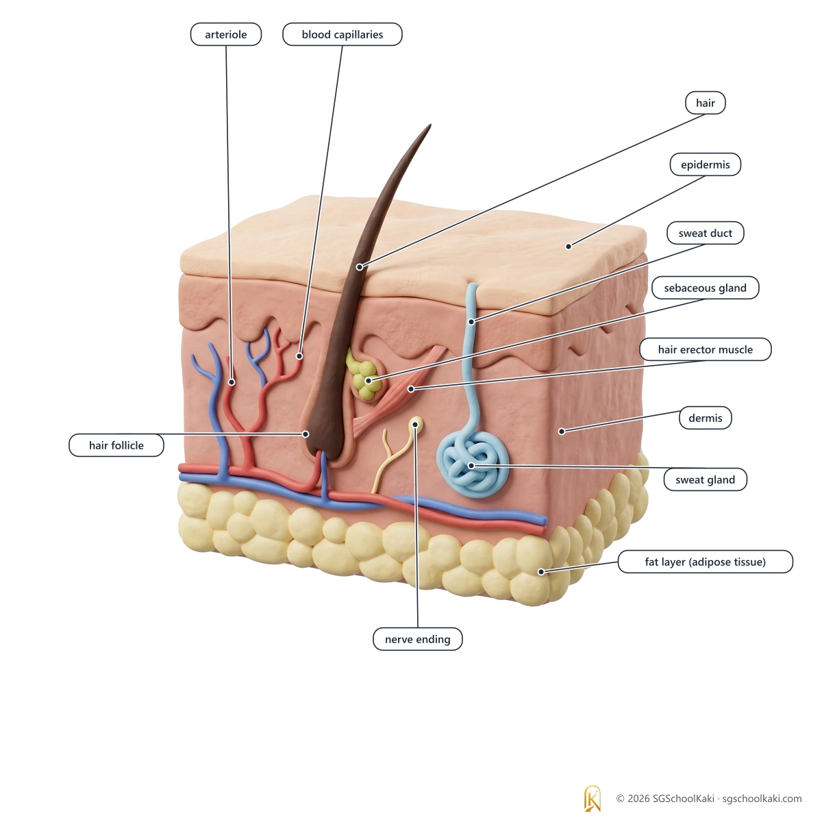
The skin: where the response happens
| Part of the skin | Job in temperature control |
|---|---|
| Nerve ending (thermoreceptor) | It detects a change in the temperature of the skin and sends impulses to the brain. |
| Sweat gland and sweat duct | The sweat gland makes sweat, and the sweat duct carries the sweat to the surface of the skin. |
| Arteriole and blood capillaries | The muscle in the wall of the arteriole changes the amount of blood that flows through the capillaries near the surface of the skin. |
| Fat cells | The layer of fat cells reduces heat loss from the body. |
| Hair and hair follicle | The hair grows from the hair follicle; the hairs of a human are short and fine, so they trap very little air and have a small effect on heat loss. |
The epidermis is the thin outer layer of the skin, the dermis below it contains the blood vessels, the sweat glands, the hair follicles and the nerve endings, and the fat cells are in a layer below the dermis. supports K325-8(c)
Sweating removes a small amount of urea from the body, so it is a form of excretion, but its main job in temperature control is to cool the body.
The temperature control loop
- 1A change in the temperature of the surroundings or of the blood is the stimulus.
- 2Thermoreceptors in the skin detect a change in the temperature of the skin and send impulses to the hypothalamus, and thermoreceptors in the hypothalamus detect the change in the temperature of the blood that flows through the brain.
- 3The hypothalamus sends impulses to the effectors, which are the sweat glands, the skeletal muscles and the muscles in the walls of the arterioles in the skin.
- 4The effectors carry out the corrective mechanisms, which bring the body temperature back to about 37 °C.
- 5The thermoreceptors detect that the set point has been restored, and the corrective mechanisms stop.
The thermoreceptors in the skin detect the temperature of the skin, which reflects the surroundings, whereas the thermoreceptors in the hypothalamus detect the temperature of the blood, which is the internal temperature.
Responses to cold and to heat
| Response | When the body is too cold | When the body is too hot | Explanation |
|---|---|---|---|
| Metabolic rate | The metabolic rate increases. | The body does not need extra heat, so the metabolic rate does not increase and may fall slightly. | Aerobic respiration in the mitochondria releases energy, and some of it is released as heat, so a higher metabolic rate produces more heat. |
| Arterioles in the skin | The arterioles constrict (vasoconstriction). | The arterioles dilate (vasodilation). | When the arterioles constrict, less blood flows through the capillaries near the surface of the skin, so less heat is lost; when they dilate, more blood flows through those capillaries, so more heat is lost. |
| Sweat glands | The sweat glands make less sweat. | The sweat glands make more sweat. | When sweat evaporates from the skin, it takes latent heat from the skin, so more sweat means more heat is lost and less sweat means less heat is lost. |
| Shivering | Shivering starts. | Shivering does not happen. | Shivering is the rapid contraction of the skeletal muscles, which increases the rate of respiration in the muscles and releases more heat. |
Shivering does not happen when the body is too hot, so you do not need to write a hot response for shivering.
Depth: why and how
- Why does sweat cool the body when it evaporates from the skin? The change from liquid to vapour takes latent heat from the skin, so the skin and the blood flowing near it lose heat.
- Why does the skin look pale on a cold day? The arterioles constrict, so less blood flows through the capillaries near the surface, and less heat is lost.
- Why does shivering warm the body? The rapid contraction of the skeletal muscles increases their rate of aerobic respiration, and some of the energy released is released as heat.
- Why is a layer of fat under the skin useful in cold weather? Fat is a poor conductor of heat, so it reduces the loss of heat from the body to the surroundings.
- Cross-link to Transport in humans: arterioles are small arteries, and the amount of blood that flows through the capillaries depends on whether the arterioles are constricted or dilated.
- Cross-link to Respiration in humans: aerobic respiration in the mitochondria releases heat, which is why a higher metabolic rate warms the body.
- Cross-link to Excretion in humans: sweat contains a small amount of urea, so sweating is a minor form of excretion.
Common mistakes
When the body is hot, the blood vessels move towards the surface of the skin.
When the body is hot, the arterioles in the skin dilate, so more blood flows through the capillaries near the surface; the blood vessels do not move.
Sweating cools the body because sweat is cold.
Sweating cools the body because the sweat takes latent heat from the skin when it evaporates.
The thermoreceptors in the skin are the control centre for body temperature.
The hypothalamus is the control centre, and the thermoreceptors in the skin detect the temperature of the skin and send impulses to the brain.
Shivering happens when the body is too hot.
Shivering happens when the body is too cold, and it does not happen when the body is too hot.
Thermoreceptors detect the change, the Hypothalamus co-ordinates, and the Arterioles, Sweat glands and Shivering respond.
The thermoreceptors in the skin and in the hypothalamus detect the change in temperature, the hypothalamus co-ordinates the response, and the effectors are the arterioles in the skin, the sweat glands and the skeletal muscles that shiver.Key: T is thermoreceptors, H is hypothalamus, A is arterioles, the first S is sweat glands and the second S is shivering.Cold: arterioles Constrict. Hot: arterioles Dilate.
When the body is too cold, the arterioles in the skin constrict so that less blood flows near the surface, and when the body is too hot, the arterioles dilate so that more blood flows near the surface.Key: The first C is cold, the second C is constrict, H is hot and D is dilate.When the body is too cold, the arterioles constrict, sweating decreases and shivering starts; when the body is too hot, the arterioles dilate, sweating increases and shivering does not happen; the hypothalamus co-ordinates the responses.
Exam check
Q1Which part of the body is the control centre for body temperature?
- ASweat gland
- BSpinal cord
- CHypothalamus
- DThermoreceptor in the skin
Show answer
MCQ answer key C (Hypothalamus): the hypothalamus receives information from the thermoreceptors and sends impulses to the effectors, so it is the control centre for body temperature.
Q2A person is in a cold room. Which response helps to keep the body temperature constant?
- AThe arterioles in the skin constrict
- BThe arterioles in the skin dilate
- CThe sweat glands make more sweat
- DShivering stops
Show answer
MCQ answer key A (The arterioles in the skin constrict): the arterioles in the skin constrict, so less blood flows through the capillaries near the surface and less heat is lost.
Q3Why does sweating lower the body temperature?
- AThe sweat takes latent heat from the skin when it evaporates
- BThe sweat is cooler than the blood and carries the heat away in the blood
- CThe sweat contains urea that absorbs heat
- DThe sweat makes the arterioles constrict
Show answer
MCQ answer key A (The sweat takes latent heat from the skin when it evaporates): the sweat takes latent heat from the skin when it evaporates, so the skin is cooled.
StructuredA student runs in hot weather and the body temperature starts to rise.
- Name the part of the brain that detects the change in the temperature of the blood. [1]
- Describe two changes in the skin that help to lower the body temperature. [2]
- Explain how shivering helps to keep the body warm in cold weather. [2]
Total: 5 marks
Show mark scheme
(a) 1 mark
- Mark scheme: hypothalamus
(b) 2 marks
- Mark scheme: the arterioles dilate, so more blood flows through the capillaries near the surface and more heat is lost
- Mark scheme: the sweat glands make more sweat, which takes latent heat from the skin as it evaporates
(c) 2 marks
- Mark scheme: the skeletal muscles contract rapidly
- Mark scheme: so the rate of respiration in the muscles increases and more heat is released
Hormones
define a hormone as a chemical substance, produced by a gland, carried by the blood, which alters the activity of one or more specific target organs
A hormone is a chemical substance, produced by a gland, carried by the blood, which alters the activity of one or more specific target organs.
- Gland
- Gland: an organ that makes a substance and releases it, such as a hormone or an enzyme supports K325-8(d)
- Target organ
- Target organ: an organ whose cells have receptors for a particular hormone, so the organ responds to that hormone supports K325-8(d)
- Chemical messenger
- Chemical messenger: a hormone carries a message from a gland to the target organs, and a very small amount of a hormone is effective supports K325-8(d)
How a hormone works
- 1A gland secretes the hormone into the blood.
- 2The blood carries the hormone around the body.
- 3The hormone reaches the target organs, whose cells have specific receptors for that hormone, and the cells of other organs do not respond to it.
- 4The hormone alters the activity of the target organs.
Hormones in this topic
| Hormone | Gland that secretes it | Target organ | Effect on the target organ |
|---|---|---|---|
| Insulin | Insulin is secreted by the islets of Langerhans in the pancreas. | The target organs of insulin are the liver and the muscles. | Insulin lowers the blood glucose concentration. |
| Glucagon | Glucagon is secreted by the islets of Langerhans in the pancreas. | The target organ of glucagon is the liver. | Glucagon raises the blood glucose concentration. |
| ADH | ADH is released by the pituitary gland. | The target organs of ADH are the kidneys. | ADH changes the amount of water that is reabsorbed by the kidneys. |
Depth: why and how
- Why do some organs respond to a hormone although the blood carries it around the whole body? The cells of the target organs have receptors for that hormone, and the other cells do not.
- Why is a hormone carried by the blood and not by nerves? The blood carries the hormone to organs in many parts of the body, so one gland can affect several target organs.
- Why is the effect of a hormone usually slower than a nerve impulse? The hormone must be secreted, carried in the blood and then bind to its receptors, which takes longer than an impulse travelling along a neurone.
- Cross-link to Transport in humans: the plasma of the blood carries hormones from the glands to the target organs.
- Cross-link to Nutrition in humans: insulin and glucagon from the pancreas control what the liver does with the glucose that is absorbed from digested food.
Common mistakes
A hormone is an enzyme made by a gland.
A hormone is a chemical substance made by a gland and carried by the blood, whereas an enzyme is a protein that acts as a biological catalyst.
Hormones travel along neurones to the target organs.
Hormones are carried by the blood, whereas nerve impulses travel along neurones.
A hormone is produced by the target organ.
A hormone is produced by a gland and alters the activity of a target organ, which is a different organ from the gland.
A hormone is a chemical substance, made by a gland, carried by the blood, and it alters the activity of specific target organs.
Exam check
Q1How is a hormone carried from a gland to the target organs?
- AAlong motor neurones
- BThrough the digestive system
- CIn the blood
- DBy diffusion through the skin
Show answer
MCQ answer key C (In the blood): a hormone is carried by the blood, whereas nerve impulses travel along neurones.
Q2Which of these is a hormone?
- AAmylase
- BInsulin
- CHaemoglobin
- DGlycogen
Show answer
MCQ answer key B (Insulin): insulin is a hormone made by the islets of Langerhans, whereas amylase is an enzyme, haemoglobin is a pigment in red blood cells and glycogen is a storage carbohydrate.
Q3The blood carries a hormone to many organs, but only some of them respond. Why?
- AThe other organs do not receive any blood
- BThe hormone is destroyed by the blood in the other organs
- CThe other organs are too far from the gland
- DThe cells of the target organs have receptors for the hormone
Show answer
MCQ answer key D (The cells of the target organs have receptors for the hormone): the cells of the target organs have specific receptors for the hormone, so the target organs respond and the other organs do not.
StructuredHormones are chemical messengers in the body.
- Define a hormone. [3]
- Name one gland that secretes a hormone, and name the hormone. [2]
- State how the hormone reaches its target organ. [1]
Total: 6 marks
Show mark scheme
(a) 3 marks
- Mark scheme: a chemical substance produced by a gland
- Mark scheme: carried by the blood
- Mark scheme: which alters the activity of one or more specific target organs
(b) 2 marks
- Mark scheme: any correct gland, for example pancreas (islets of Langerhans), OR pituitary gland, OR adrenal gland, OR testes, OR ovaries
- Mark scheme: the matching hormone, for example insulin or glucagon for the pancreas, OR ADH for the pituitary gland, OR adrenaline for the adrenal glands, OR testosterone for the testes, OR oestrogen for the ovaries
(c) 1 mark
- Mark scheme: it is carried by the blood (in the plasma)
Endocrine glands and the islets of Langerhans
explain what is meant by an endocrine gland, with reference to the islets of Langerhans in the pancreas
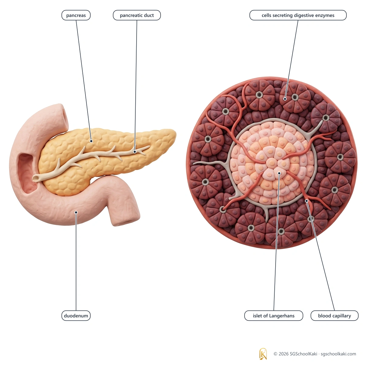
An endocrine gland is a ductless gland that secretes its hormones directly into the blood, and the islets of Langerhans in the pancreas are an example.
- Endocrine gland
- Endocrine gland: a gland with no duct, which secretes hormones directly into the blood
- Exocrine gland
- Exocrine gland: a gland that secretes its product into a duct, such as a salivary gland or a sweat gland supports K325-8(e)
- Pancreas
- Pancreas: an organ that has an exocrine part, which makes pancreatic juice that flows through the pancreatic duct into the duodenum, and an endocrine part, the islets of Langerhans supports K325-8(e)
- Islets of Langerhans
- Islets of Langerhans: groups of cells scattered through the pancreas, which secrete the hormones insulin and glucagon directly into the blood
Endocrine and exocrine glands
| Feature | Endocrine gland | Exocrine gland |
|---|---|---|
| Duct | An endocrine gland has no duct. | An exocrine gland has a duct. |
| Where the product goes | An endocrine gland secretes its product directly into the blood. | An exocrine gland secretes its product through a duct onto a surface or into a cavity. |
| Product | The product of an endocrine gland is a hormone. | The product of an exocrine gland is, for example, an enzyme, saliva, sweat or mucus. |
| Examples | The islets of Langerhans in the pancreas and the pituitary gland are endocrine glands. | The salivary glands, the sweat glands and the part of the pancreas that makes pancreatic juice are exocrine glands. |
Other endocrine glands include the pituitary gland, the thyroid gland, the adrenal glands, the ovaries and the testes. supports K325-8(e)
Two different types of cell in the islets of Langerhans secrete the two hormones, so one islet makes both insulin and glucagon.
The beta cells of the islets secrete insulin, and the alpha cells of the islets secrete glucagon. supports K325-8(e)
Depth: why and how
- Why does an endocrine gland have a rich blood supply? The hormones are secreted directly into the blood, so the gland needs many capillaries close to its cells to pick up the hormone.
- Why does the pancreas count as both an endocrine gland and an exocrine gland? The islets of Langerhans secrete hormones into the blood, whereas the rest of the pancreas secretes pancreatic juice into a duct.
- Cross-link to Nutrition in humans: the pancreatic juice from the exocrine part of the pancreas contains enzymes that digest carbohydrate, protein and fat in the small intestine.
- Cross-link to Transport in humans: the blood plasma carries the hormones from the endocrine glands to the target organs.
Insulin and glucagon are proteins, so insulin would be digested if it were swallowed, which is why it is given by injection. supports K325-8(e)
Common mistakes
An endocrine gland releases its hormones through a duct.
An endocrine gland is ductless: it releases its hormones directly into the blood.
The pancreas is an endocrine gland and nothing else.
The pancreas has an endocrine part, the islets of Langerhans, and an exocrine part that secretes pancreatic juice into the pancreatic duct.
Insulin and glucagon are made by the same type of cell in the islets.
Two different types of cell in the islets make the two hormones: beta cells make insulin and alpha cells make glucagon.
Endocrine means ductless: the islets of Langerhans in the pancreas secrete insulin and glucagon directly into the blood.
Exam check
Q1What is an endocrine gland?
- AA gland that secretes enzymes into a duct
- BA gland that secretes hormones into a duct that leads to an organ
- CA gland that secretes hormones directly into the blood
- DA gland that secretes sweat onto the surface of the skin
Show answer
MCQ answer key C (A gland that secretes hormones directly into the blood): an endocrine gland has no duct and secretes its hormones directly into the blood.
Q2Which structures in the pancreas secrete insulin and glucagon?
- AIslets of Langerhans
- BPancreatic ducts
- CVilli
- DSalivary glands
Show answer
MCQ answer key A (Islets of Langerhans): the islets of Langerhans are groups of cells in the pancreas that secrete insulin and glucagon directly into the blood.
Q3Which statement about the pancreas is correct?
- AIt secretes both hormones and digestive enzymes into the blood
- BIt secretes both hormones and digestive enzymes into a duct
- CIt secretes digestive enzymes into the blood and hormones into a duct
- DIt secretes hormones into the blood and digestive enzymes into a duct
Show answer
MCQ answer key D (It secretes hormones into the blood and digestive enzymes into a duct): the islets of Langerhans secrete hormones into the blood, and the exocrine part secretes pancreatic juice, which contains digestive enzymes, into the pancreatic duct.
StructuredThe islets of Langerhans are found in the pancreas.
- Name the two hormones that are secreted by the islets of Langerhans. [2]
- Explain why the islets of Langerhans are called an endocrine gland. [2]
- State one difference between an endocrine gland and an exocrine gland. [1]
Total: 5 marks
Show mark scheme
(a) 2 marks
- Mark scheme: insulin
- Mark scheme: glucagon
(b) 2 marks
- Mark scheme: they secrete hormones
- Mark scheme: directly into the blood, because they have no duct
(c) 1 mark
- Mark scheme: an endocrine gland has no duct and an exocrine gland has a duct, OR an endocrine gland secretes into the blood and an exocrine gland secretes into a duct
Blood glucose: insulin and glucagon
explain how blood glucose concentration is regulated by insulin and glucagon as a homeostatic mechanism
The blood glucose concentration is kept constant by negative feedback using two hormones from the islets of Langerhans: insulin lowers the blood glucose concentration and glucagon raises it.
- Blood glucose concentration
- Blood glucose concentration: the amount of glucose dissolved in the blood, which is kept within narrow limits around the set point supports K325-8(f)
- Insulin
- Insulin: a hormone secreted by the islets of Langerhans when the blood glucose concentration is high, which lowers the blood glucose concentration
- Glucagon
- Glucagon: a hormone secreted by the islets of Langerhans when the blood glucose concentration is low, which raises the blood glucose concentration
- Glycogen
- Glycogen: a storage carbohydrate made of many glucose molecules joined together, which is stored in the liver and the muscles supports K325-8(f)
Insulin and glucagon side by side
| Feature | Insulin | Glucagon |
|---|---|---|
| Stimulus | The stimulus for insulin is a high blood glucose concentration. | The stimulus for glucagon is a low blood glucose concentration. |
| Detected and secreted by | The cells in the islets of Langerhans in the pancreas detect the stimulus and secrete insulin. | The cells in the islets of Langerhans in the pancreas detect the stimulus and secrete glucagon. |
| Target organs | The target organs of insulin are the liver and the muscles. | The target organ of glucagon is the liver. |
| Response of the target cells | Insulin makes the liver cells and the muscle cells take up more glucose, and glucose is converted to glycogen. | Glucagon causes the liver cells to convert glycogen to glucose, which is released into the blood. |
| Effect on the blood glucose concentration | Insulin lowers the blood glucose concentration to the set point. | Glucagon raises the blood glucose concentration to the set point. |
Glucagon acts on the liver and not on the muscles, because the glycogen in the muscles is used by the muscle cells themselves and is not released as glucose into the blood.
Two loops, one set point
When the blood glucose concentration is high
- 1After a meal, glucose from the digested carbohydrate is absorbed into the blood, and the blood glucose concentration rises above the set point, which is the stimulus.
- 2The cells in the islets of Langerhans detect the high blood glucose concentration and secrete insulin into the blood.
- 3The blood carries the insulin to the liver and the muscles, which are the target organs.
- 4Insulin makes the liver cells and the muscle cells take up more glucose, and glucose is converted to glycogen; for SEAB answers, write that insulin increases the permeability of the cells to glucose.
- 5The blood glucose concentration falls to the set point, the islet cells detect this and less insulin is secreted, so the corrective mechanism stops.
When the blood glucose concentration is low
- 1Between meals and during exercise, the cells use glucose in respiration, and the blood glucose concentration falls below the set point, which is the stimulus.
- 2The cells in the islets of Langerhans detect the low blood glucose concentration and secrete glucagon into the blood.
- 3The blood carries the glucagon to the liver, which is the target organ.
- 4Glucagon causes the liver cells to convert glycogen to glucose, which is released into the blood.
- 5The blood glucose concentration rises to the set point, the islet cells detect this and less glucagon is secreted, so the corrective mechanism stops.
Glycogen is a carbohydrate that is stored in the liver and the muscles, whereas glucagon is a hormone that is secreted by the islets of Langerhans.
Depth: why and how
- Why is glucose stored as glycogen and not as glucose? Glycogen is insoluble, so it does not change the water potential of the cells, whereas a large amount of dissolved glucose would.
- Why do two hormones with opposite effects give better control than one? The blood glucose concentration can be pushed back up when it falls and pulled back down when it rises, so it stays close to the set point.
- Why is the liver the main target organ for both hormones? It stores a large amount of glycogen, and the hepatic portal vein brings blood that contains the absorbed glucose from the small intestine directly to the liver.
- Cross-link to Nutrition in humans: glucose from the digestion of carbohydrate is absorbed into the capillaries of the villi and reaches the liver in the hepatic portal vein.
- Cross-link to Transport in humans: insulin and glucagon are carried in the blood plasma to the liver and the muscles.
- Cross-link to Respiration in humans: the blood glucose concentration falls during exercise because the muscles use glucose for respiration, so glucagon is secreted.
Common mistakes
Insulin changes glycogen to glucose.
Insulin causes glucose to be converted to glycogen, and glucagon causes glycogen to be converted to glucose.
Glucagon acts on the liver and the muscles.
Glucagon acts on the liver, whereas insulin acts on the liver and the muscles.
Insulin is secreted when the blood glucose concentration is low.
Insulin is secreted when the blood glucose concentration is high, and glucagon is secreted when it is low.
Glycogen is the hormone that raises the blood glucose concentration.
Glucagon is the hormone that raises the blood glucose concentration, and glycogen is the carbohydrate that is stored in the liver and the muscles.
Insulin lets glucose IN to the liver and muscle cells; Glucagon lets glucose GO out of the liver, not out of the muscles.
Insulin makes the liver cells and the muscle cells take glucose in and store it as glycogen, whereas glucagon makes the liver, but not the muscles, release glucose into the blood.Key: The first I is insulin, the second I is in, the first G is glucagon and the second G is go.Glycogen is the Store; Glucagon is the Signal that empties it.
Glycogen is the storage form of glucose in the liver and the muscles, whereas glucagon is the hormone that signals the liver to change glycogen to glucose.Key: The first G is glycogen, the first S is store, the second G is glucagon and the second S is signal.High blood glucose: insulin acts on the liver and muscles and glucose becomes glycogen. Low blood glucose: glucagon acts on the liver and glycogen becomes glucose.
Exam check
Q1Which hormone is secreted when the blood glucose concentration is high?
- AGlucagon
- BInsulin
- CGlycogen
- DAmylase
Show answer
MCQ answer key B (Insulin): insulin is secreted when the blood glucose concentration is high, whereas glucagon is secreted when it is low.
Q2Which organs does insulin act on to lower the blood glucose concentration?
- AThe liver only
- BThe liver and the muscles
- CThe muscles and the kidneys
- DThe pancreas and the stomach
Show answer
MCQ answer key B (The liver and the muscles): the target organs of insulin are the liver and the muscles, where glucose is converted to glycogen.
Q3What does glucagon cause the liver to do?
- AConvert glucose to glycogen
- BMake more insulin
- CIncrease the permeability of the muscle cells to glucose
- DConvert glycogen to glucose and release it into the blood
Show answer
MCQ answer key D (Convert glycogen to glucose and release it into the blood): glucagon causes the liver cells to convert glycogen to glucose, which is released into the blood, so the blood glucose concentration rises.
StructuredAfter a meal that is rich in carbohydrate, the blood glucose concentration rises.
- Name the hormone that is secreted and the part of the pancreas that secretes it. [2]
- Describe how this hormone lowers the blood glucose concentration. [3]
- Explain how the blood glucose concentration returns to the set point when it falls below normal between meals. [3]
Total: 8 marks
Show mark scheme
(a) 2 marks
- Mark scheme: insulin
- Mark scheme: islets of Langerhans
(b) 3 marks
- Mark scheme: it is carried by the blood to the liver and the muscles
- Mark scheme: it makes the liver cells and the muscle cells take up more glucose, OR it increases the permeability of the cells to glucose
- Mark scheme: glucose is converted to glycogen
(c) 3 marks
- Mark scheme: the islets of Langerhans detect the low blood glucose concentration and secrete glucagon
- Mark scheme: glucagon acts on the liver, which converts glycogen to glucose
- Mark scheme: the glucose is released into the blood, so the blood glucose concentration rises to the set point
You've read the free 34%. Unlock the rest of Topic 8.
Still to read in this topic:
- Type 2 diabetes mellitus
- Risk factors and management of type 2 diabetes
- ADH and osmoregulation
- The nervous system
- Neurones and reflex action
- Structure of the eye
- Focusing on near and distant objects
- The pupil reflex
$9 one-time · all 14 topics · no subscription
Less than a fast-food meal.
Yours for the whole O-Level.
Premium members get all 14 topics included.
Notes version 1.0 · Last updated 30 Sept 2026 · Topic 8 of 14